معلومات ذات صله

استشر الولايات المتحدة
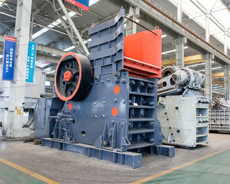
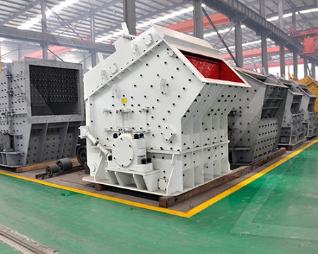
بصفتنا مصنعًا عالميًا رائدًا لمعدات التكسير والطحن ، فإننا نقدم حلولًا متطورة وعقلانية لأي متطلبات لتقليل الحجم ، بما في ذلك إنتاج المحاجر والركام والطحن ومحطة تكسير الحجارة الكاملة. نقوم أيضًا بتوريد الكسارات والمطاحن الفردية وكذلك قطع غيارها.


Manganeseenhanced magnetic resonance imaging (MEMRI)
Manganeseenhanced MRI (MEMRI) evolved in the late nineties when Koretsky and associates pioneered the use of MEMRI for brain activity measurements (2)MEMRI relies upon the following three main properties of Mn 2+: (1) it is a paramagnetic ion that shortens the spin lattice relaxation time constant ( T1) of tissues, where itManganeseEnhanced Magnetic Resonance Imaging (MEMRI)
Manganeseenhanced magnetic resonance imaging (MEMRI)
2013年10月2日· An ex vivo functional imaging technique, manganeseenhanced magnetic resonance imaging (MEMRI), was used to identify and distinguish brain regions that2020年2月25日· Abstract Manganeseenhanced magnetic resonance imaging (MEMRI) relies on the strong paramagnetism of Mn2+ Mn2+is a calcium ion analog and can enterManganeseEnhanced Magnetic Resonance Imaging: Application
ManganeseEnhanced Magnetic Resonance Imaging: Overview
2018年12月13日· Manganeseenhanced magnetic resonance imaging (MEMRI) rose to prominence in the 1990s as a sensitive approach to high contrast imaging Following theThe metal manganese is a potent magnetic resonance imaging (MRI) contrast agent that is essential in cell biology Manganeseenhanced magnetic resonance imagingManganeseenhanced MRI: an exceptional tool in translational
Manganeseenhanced magnetic resonance imaging (MEMRI
Manganeseenhanced magnetic resonance imaging (MEMRI): methodological and practical considerations 2004 Dec;17 (8):53243 doi: 101002/nbm945 Afonso C Silva2023年11月1日· Manganeseenhanced magnetic resonance assessment of changes in hippocampal neural function after the treatment of radiationinduced brain injury withManganeseenhanced magnetic resonance assessment of changes
ManganeseEnhanced Magnetic Resonance Imaging:
2020年2月25日· Manganeseenhanced magnetic resonance imaging (MEMRI) relies on the strong paramagnetism of Mn 2+Mn 2+ is a calcium ion analog and can enter excitable cells through voltagegated calcium channels Mn 2+ can be transported along the axons of neurons via microtubulebased fast axonal transport Based on these properties, MEMRIThe metal manganese is a potent magnetic resonance imaging (MRI) contrast agent that is essential in cell biology Manganeseenhanced magnetic resonance imaging (MEMRI) is providing unique information in an evergrowing number of applications aimed at understanding the anatomy, the integration, and the function of neural circuits both inManganeseenhanced MRI: an exceptional tool in translational
ManganeseEnhanced Magnetic Resonance Imaging (MEMRI)
The use of manganese ions (Mn 2+) as an MRI contrast agent was introduced over 20 years ago in studies of Mn 2+ toxicity in anesthetized rats (1) Manganeseenhanced MRI (MEMRI) evolved in the late nineties when Koretsky and associates pioneered the use of MEMRI for brain activity measurements (2) as well as neuronal tract tracing (3)Manganeseenhanced magnetic resonance imaging of mouse brain after systemic administration of MnCl2: dosedependent and temporal evolution of T1 contrast (9175 mg/kg) were administered to mice from 0 to 24 h prior to T(1)weighted manganeseenhanced MRI (MEMRI) at 117 T PreMnCl(2) T(1) values measured in different brainManganeseenhanced magnetic resonance imaging of mouse brain
Manganeseenhanced magnetic resonance imaging (MEMRI) of rat brain
2011年1月1日· Manganese (Mn(2+))enhanced magnetic resonance (MR) imaging (MEMRI) in rodents offers unique opportunities for the longitudinal study of hippocampal structure and function in parallel with cognitive testing However, Mn(2+) is a potent toxin and there is evidence that it can interfere with neuronalManganese ion (Mn2+) is an essential metal that participates as a cofactor in a number of critical biological functions, such as electron transport, detoxification of free radicals and synthesis of neurotransmitters Mn2+ can enter excitable cells using some of the same transport systems as Ca2+ andManganeseenhanced magnetic resonance imaging (MEMRI)
ManganeseEnhanced MRI: Biological Applications in Neuroscience
2015年7月10日· Magnetic resonance imaging (MRI) is an excellent noninvasive tool to investigate biological systems The administration of the paramagnetic divalent ion manganese (Mn 2+) enhances MRI contrast in vivoDue to similarities between Mn 2+ and calcium (Ca 2+), the premise of manganeseenhanced MRI (MEMRI) is that the former2022年8月8日· In addition to applications in anatomical T2weighted magnetic resonance imaging (MRI), we also describe applications in acquiring 31P spectra, manganeseenhanced magnetic resonance imaging (MEMRI) transport rates and restingstate functional magnetic resonance imaging (rsfMRI) in awake animals and describe aA Mouse Holder for Awake Functional Imaging in Unanesthetized
In vivo, transsynaptic tracttracing utilizing manganeseenhanced
Manganeseenhanced MRI (MEMRI) utilizes a combination of these properties of Mn2+ to trace neuronal pathways in an MRIdetectable manner The focus of this review will detail some of the current MEMRI tracttracing methodologies in mice and nonhuman primates as well as biological applications of MEMRI tracttracing2004年11月1日· To this end, we now report that manganese (Mn(2+))enhanced magnetic resonance imaging (MEMRI) can reflect brain regionspecific HIV1induced neuropathology in chronically virusinfected NOD/scidManganeseenhanced magnetic resonance imaging (MEMRI) of brain
Manganeseenhanced magnetic resonance imaging PubMed
DOI: 101007/978161779219928 Manganeseenhanced magnetic resonance imaging (MEMRI) relies on contrasts that are due to the shortening of the T (1) relaxation time of tissue water protons that become exposed to paramagnetic manganese ions In experimental animals, the technique combines the high spatial resolution achievable by2018年4月28日· Manganeseenhanced magnetic resonance imaging (MEMRI) rose to prominence in the 1990s as a sensitive approach to high contrast imaging Brain magnetic resonance imaging with contrast dependent on blood oxygenation Proc Natl Acad Sci U S A 87, 9868–9872 101073/pnas87249868 PMID: Manganeseenhanced magnetic resonance imaging: Overview
ManganeseEnhanced Magnetic Resonance Imaging (MEMRI)
Indeed, manganeseenhanced MRI (MEMRI) has been gaining growing interest in the past few years ( 2, 3, 36, 37) and currently has three main applications for biological systems First, owing to its contrastenhancing properties, systemic Mn 2+ injections are used for enhancement of the brain cytoarchitecture for anatomical studies ( 38 – 44 )2023年10月18日· In this study, we aimed to evaluate the effects of BMSCs treatment on changes in hippocampal neural function in SpragueDawley(SD) rats with RIBI, and to evaluate the therapeutic effect of BMSCs by manganeseenhanced magnetic resonance imaging (MEMRI) First, we assessed cognitive function after RIBI treatment withManganeseenhanced magnetic resonance assessment of
ScienceDirect Postmortem neuroimaging: Temporal and spatial
2021年8月1日· Recent studies have addressed these concerns by using manganeseenhanced magnetic resonance imaging (MEMRI) Basic challenges for in vivo application of MEMRI in rodents includes how to set inclusion criteria for adequate Mn 2+ uptake and whether valid data can be collected from brains postmortem2013年10月2日· One tool for identifying activated neuronal networks is manganeseenhanced magnetic resonance imaging (MEMRI) [16–18] Unlike imaging based on blood oxygenation or blood flow, MEMRI is based on the ability of paramagnetic manganese ions (Mn 2+ ) to enter activated neurons through voltagegated calcium channels [ 16 ,Manganeseenhanced magnetic resonance imaging (MEMRI) reveals brain
Applications of ManganeseEnhanced Magnetic Resonance Imaging
2019年5月14日· As such, development of imaging techniques for the visual system is crucial for unveiling the neural basis of visual function or impairment Magnetic resonance imaging (MRI) offers noninvasive probing of the structure and function of the neural circuits without depth limitation, and can help identify abnormalities in brain tissues in vivo2022年12月1日· This study aimed to determine whether manganeseenhanced magnetic resonance imaging (MEMRI) could be used to assess stress in rat brains Methods We exposed male Wistar/ST rats bred in a specific pathogenfree environment to ultrasound stimuli (22 kHz and 55 kHz) for 10 days and then assessed brain activities usingManganeseenhanced magnetic resonance imaging detects
Manganeseenhanced magnetic resonance imaging (MEMRI)
The MEMRI neuronal activity studies have been generally performed either by stereotactic brain injection or by systemic in conjunction with t Manganeseenhanced magnetic resonance imaging (MEMRI) without compromise of the bloodbrain barrier detects hypothalamic neuronal activity in vivo NMR Biomed 2006 Dec;19(8):年1月1日· Manganeseenhanced magnetic resonance imaging (MEMRI) relies on contrasts that are due to the shortening of the T 1 relaxation time of tissue water protons that become exposed to paramagnetic manganese ions In experimental animals, the technique combines the high spatial resolution achievable by MRI with the biological informationManganeseEnhanced Magnetic Resonance Imaging